OncoWuXi Express: In Vitro Hydrogel-based 3D Tumor Models
Introduction:
Over the last few years, the clinical success of immune checkpoint inhibitors has increasingly drawn attention to cancer therapies that target the tumor microenvironment (TME). TME consists of various cell types that are embedded in the extracellular matrix (ECM), including immune cells, endothelial cells, and cancer associated fibroblasts, all of which play critical roles in drug resistance and tumor recurrence [1]. Therefore, incorporating diverse elements of TME can enhance the predictive power of in vitro tumor models, improve the precision of drug screening, and provide a broader range of options for investigating interactions between tumor cells and TME.
Traditional in vitro models, like the 2D cell culture system and simple spheroid culture system, often fall short with in vivo translatability due to their lack of TME elements. This absence hampers their ability to accurately depict the complexity of in vivo tumors, posing challenges for early-stage drug screening. To overcome this limitation, we developed VitroGel®-based tumor models to mimic the natural extracellular matrix environment, enabling the co-culture of tumor cells and other cell types within an in vitro 3D hydrogel system. This model can also be established in standard 96-well plate format, facilitating high-throughput cell viability and cytotoxicity screening assays. Moreover, WuXi Biology offers a customized hydrogel-based 3D tumor model service, allowing for faster, more reliable, and personalized solutions for new drug development and drug mechanism exploration.
1. In vitro growth monitoring of hydrogel-based 3D tumor models
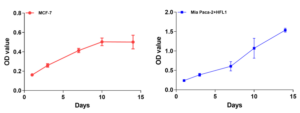
MCF-7 cells were encapsulated solely within the hydrogel matrix for 3D cell culture. MIA PaCa-2 cells and human fetal lung fibroblast 1 cells (HFL1) were encapsulated in the hydrogel matrix for 3D cell co-culture. The growth curves below were measured by the CCK8 kit (Figure 1).

Figure 1. Growth curve of hydrogel-based 3D MCF-7 tumor model (Left), growth curve of hydrogel-based 3D MIA PaCa-2 and HFL1 co-culture tumor model (Right)
2. In vitro efficacy evaluation in hydrogel-based 3D tumor models
To assess the effects of TME elements on efficacy evaluation, we established 3D tumor models by encapsulating NCI-H358 cells, with or without HFL1 cells, in the hydrogel matrix within 96-well plates. Subsequently, the cell culture was treated with docetaxel for 5 days. Cell images were captured by a microscope and Incucyte system. The anti-tumor effect of docetaxel was evaluated by CTG assay. The results showed that co-culturing HFL1 with NCI-H358 cells produced a stronger response to docetaxel.

Figure 2. Representative images of hydrogel-based 3D NCI-H358 and HFL1 co-culture tumor model (Left), anti-tumor effect of docetaxel on hydrogel-based 3D NCI-H358 tumor model with or without HFL1 cells (Right).
3. In vitro viability analysis of hydrogel-based 3D tumor models
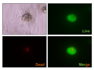
AMG 510-resistant MIA PaCa-2 (AMG 510-R-MIA PaCa-2) cells were co-cultured with HFL1 cells in the hydrogel matrix under 3D conditions, following a 10-day treatment with AMG 510. Subsequently, Cyto3D reagent was added for cell viability analysis.

Figure 3. Live-dead cell viability images of hydrogel-based 3D AMG 510-R-MIA PaCa-2 and HFL1 co-culture tumor model. Green: live cells. Red: dead cells.
Conclusion:
Hydrogel-based 3D tumor models can effectively replicate the morphology and environment of solid tumors. This enhances the accuracy of drug screening, providing strong support for exploring mechanisms and developing cancer therapies based on tumor-stroma interactions.
Our hydrogel-based 3D tumor models and services include:
- Hydrogel-based 3D tumor models (tumor cells alone)
- Hydrogel-based 3D co-culture tumor models (tumor cells + other cell types)
- Growth curves
- Cell viability/ cytotoxicity assays
- Integration with other analyses (e.g., WB, qPCR)
References:
[1] Xu Maosen, Zhang Tao, Xia Ruolan et al. Targeting the tumor stroma for cancer therapy. J. Mol Cancer, 2022, 21: 208.
WuXi AppTec | Oncology Platform:
Related Content
Despite the success of delivery technologies such as lipid nanoparticles and trivalent GalNAc conjugation, targeted delivery of oligonucleotides to extra...
VIEW RESOURCEWuXi AppTec scientists contributed to a research article in the journal Translational Medicine Communications which characterized the antitumor immune responses...
VIEW RESOURCE
