
Analysis of Cellular and Tissue Biomarkers in NHP Samples
OncoWuXi Express will continue to keep you informed about updates to our online pharmacology model database (OncoWuXi Database), as well as our recent progress in preclinical research. In this issue, we showcase the analysis of cellular and tissue biomarkers in NHP samples.
![]()
https://onco.wuxiapptec.com
Introduction:
Non-human primates (NHPs) are highly similar to humans in terms of genetics, immune systems, and physiological structures, making them an important model for studying human diseases and evaluating drug efficacy. With the rapid advancement of cutting-edge research areas such as immunotherapy and neurological disorders, there is a growing demand for high-precision flow cytometry analysis and histopathological evaluation using NHP tissue samples.
WuXi Biology has successfully established and systematically validated a standardized flow cytometry and histopathological analysis system for multiple tissue samples from cynomolgus monkeys. The core technical advantages of this tissue flow cytometry platform are as follows:
- Rapid Sampling: A standardized process for tissue sampling
- Precise Enzymatic Digestion: Optimized enzymatic digestion protocols tailored for different tissues (Figure 1A), ensuring the acquisition of highly viable single-cell suspensions (Figure 1B).
- Cross-Validation: Compatibility challenges of NHP-specific antibodies have been overcome, with cross-validation for multiple antibodies.
- Broad Spectrum Analysis: full-spectrum flow cytometer that supports multi-parameter panel analysis of up to 21 colors (Figure 1C).
Through this tissue flow cytometry platform, it is possible to comprehensively cover key preclinical research scenarios, including but not limited to detailed immune cell subset profiling, mechanism studies of neurological diseases, and drug tissue distribution analysis.
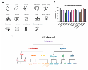
Figure 1. Overview of tissue flow cytometry analysis in cynomolgus monkeys
Case Study
Case 1: Liver Tissue
WuXi Biology has established a wedge-shaped liver biopsy technique in the NHP model, enabling the acquisition of sufficient liver tissue samples under non-terminal conditions for multidimensional analysis. Based on this technical platform, the team further integrated flow cytometry with histopathological methods to systematically evaluate the liver’s immune microenvironment and cellular composition.
In terms of flow cytometry analysis, WuXi Biology developed efficient enzymatic dissociation protocols and specific antibody detection panels tailored to immune cells and parenchymal/non-parenchymal liver cells. These methods enable the precise quantification of the phenotypic characteristics, activation, and proliferation states of immune subsets such as T cells, B cells, and NK cells. Additionally, they enable the classification and functional analysis of parenchymal cells (hepatocytes) and non-parenchymal cells (e.g., Kupffer cells) (Figure 2A).
Moreover, WuXi Biology established a multiplex immunofluorescence (mIF) detection system based on single samples, which can simultaneously display the spatial distribution of three cell types in the same liver tissue slice. This approach, combined with advanced image analysis software, provides high-precision quantification (Figure 2B). This technological platform offers robust support for research into drug distribution in liver tissues and the immune microenvironment.
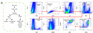
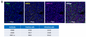
Figure 2. Flow cytometry analysis and multiplex immunofluorescence staining of liver tissue in cynomolgus monkeys
Case Study
Case 2: Brain Tissue
WuXi Biology has established a comprehensive neuroanatomical technical system, enabling precise dissection and partitioning of brain region tissues. Leveraging this platform, we have successfully developed an enzymatic digestion-flow cytometry combined analysis method for specific brain tissues, allowing the quantitative detection of immune cell subsets (Figure 3A&B) [1].
In terms of histopathological analysis, WuXi Biology utilized immunofluorescence labeling techniques to achieve precise localization analysis of various subdivided brain regions. This study specifically highlights the spatial expression characteristics of the vascular endothelial cell marker CD31 in different functional partitions (Figure 3C).


Figure 3. Flow cytometry analysis and immunofluorescence staining of brain tissue in cynomolgus monkeys
Case Study
Case 3: Bone Marrow Tissue
WuXi Biology has developed a non-terminal bone marrow aspiration technique, enabling dynamic and repeated collection of bone marrow samples in the cynomolgus monkey model. Utilizing this platform, the team has established a multidimensional bone marrow cell analysis system centered on flow cytometry. This system covers immune phenotyping, B cell depletion levels, and quantitative detection of hematopoietic stem cells (HSCs). Figure 4A demonstrates the gating strategy and detection results for HSCs via flow cytometry.
In terms of bone marrow histopathological analysis, WuXi Biology has standardized the fixation and HE staining procedures for bone marrow aspirate clots [2] and developed IHC staining protocols for multiple biomarker targets (Figure 4B). These advancements provide robust histological support for the dynamic monitoring of the hematopoietic microenvironment and pharmacodynamic changes.

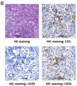
Figure 4. Flow cytometry analysis and histological staining of bone marrow tissue in cynomolgus monkeys
Summary
In NHP tissue analysis, flow cytometry and pathological analysis complement each other. Flow cytometry reveals dynamic and functional changes at the cellular level, while pathological analysis provides tissue localization information. Together, they drive the translation from basic research to clinical applications and serve as essential tools for studying disease mechanisms and evaluating pharmacodynamics.
References:
[1] Croese, T., Castellani, G., & Schwartz, M. (2021). Immune cell compartmentalization for brain surveillance and protection. Nature immunology, 22(9), 1083-1092.
[2] Salamanna, F., Contartese, D., Nicoli Aldini, N., Barbanti Brodano, G., Griffoni, C., Gasbarrini, A., & Fini, M. (2018). Bone marrow aspirate clot: A technical complication or a smart approach for musculoskeletal tissue regeneration. Journal of Cellular Physiology, 233(4), 2723-2732.
If you have any questions or need further information, please contact us at:
Business Consulting:
Technical Contact:
Pharmacology-BD-Translation@wuxiapptec.com
Scan the QR code below to register at our OncoWuXi database for free.
For more pharmacological model data, information, and consultation.

![]()
https://onco.wuxiapptec.com
Related Content
Receptor tyrosine kinase-like orphan receptor 1 (ROR1) is a key mediator of non-canonical Wnt signaling and plays a critical role...
VIEW RESOURCEAt this year’s annual meeting of the American Association for Cancer Research (AACR), WuXi Biology has been selected to present...
VIEW RESOURCE