Histology & Histopathology Labs
Recognized for our breadth of capabilities and professional expertise in in vivo pharmacology and biomarker discovery, WuXi Biology merges our talents in histopathology to provide an advanced support to preclinical drug discovery. We provide services in histology, toxicological pathology, and disease pathology such as oncology, inflammation, metabolic diseases and neurodegeneration with extensive and comprehensive technology and expertise and pathology evaluation. The pathology team offers a wide range of preclinical histology and pathology services spanning all aspects of paraffin and frozen slide preparation, staining, and evaluation at the designated histology labs and closely works with our clients to assure the highest quality on our histopathology studies. This combination of experience and space affords excellent capacity and throughput to meet clients’ timelines.
Capability Overview
- Histology laboratory capability
- Special histochemistry
- Immunohistochemistry
- Disease pathology
- Toxicologic pathology
- Histomorphometry
- Tissus banks of rodent, rabbit, dog monkey and human
- Interpretation via board-certified pathologists and slide review over th internet
Histopathology lab skills
- Small and large animal necropsy
- In-situ perfusion fixation
- Ex-vivo organ perfusion fixation
- Micro-dissection
- High resolution gross necropsy photography
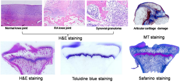
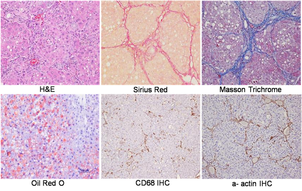
- Regular H&E staining
- Special staining: PAS, Masson trichrome, Silver, PAM, Sirius red, Oil red Sudan III and other requested special staining
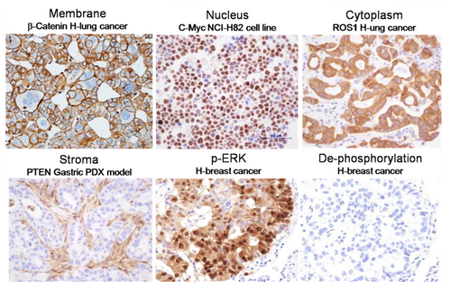
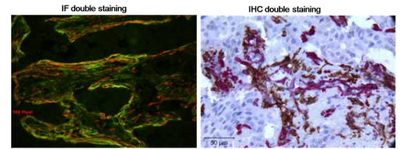
- Immunohistochemical staining (IHC)
- Immunofluorescent staining (IF)
- Immunocytochemical staining (ICC)
- In situ hybridization (ISH)
- Fluorescent in situ hybridization (FISH)
Experimental pathological evaluation capabilities
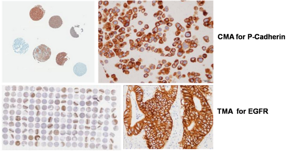
- Analysis for oncology study such as biomarker screen, location, density on cell lines, CDX, tissue microarray (TMA), cell line microarray (CMA)
- Analysis for inflammatory / immune disease models such as EAE model, IBD model, lung fibrosis model, COPD model and CIA model
- Analysis for tissue and organ injuries such as stroke, AMI, kidney diseases, pain, CNS diseases
- Analysis for tissue and organ metabolic and degeneration such as liver diseases (NASH, NAFLD), skeleton muscle degeneration, bone degeneration, and connective tissue degeneration
Toxicological pathology
Non-GLP toxic pathology including gross pathology and histopathology reading, analysis, evaluation, review and report
Biomarker assay development and validation
Biomarker reorganization, localization, screening via IHC, FISH, ISH on TMA, CMA, human tissue bank, animal tissue bank as well NHP tissue bank
Equipment and Facility
- Organ perfusion tanks
- Gross necropsy photography station
- Light microscopes with digital photography
- Automatic histoprocessor
- Automatic stainer
- Paraffin microtomes
- Fluorescent microscopes with digital photography
- Cryostat sectioning
- -80 C degree tissue sample storage
- Liquid nitrogen tissue sample storage
- Regular paraffin embedded block sample storage
- Biosafety hood