
In Vivo Pharmacology Services for Ophthalmic Drug Development
Introduction
The number of people with vision impairment has increased due to the continuously expanding and aging population, as well as lifestyle changes. According to the Lancet Global Health, by 2050, myopia is expected to affect 5 billion people. Additionally, the number of individuals with distance vision impairment is projected to reach 895 million, including 61 million who will be blind worldwide [1].

Figure 1: The human eye and common conditions [1]
Currently, there is a significant burden of ophthalmic diseases such as cataracts, glaucoma, and age-related macular degeneration (AMD), leading to an increased need for prevention and treatment strategies (Figure 2). AMD, in particular, is a leading cause of irreversible vision loss in the elderly and can be classified into dry (atrophic) and wet (exudative) forms. Dry AMD typically starts with early deposit formation (drusen) in the retina, progresses to retinal pigment epithelium degeneration, and then culminates in the death of photoreceptor cells (retinal atrophy), ultimately resulting in vision loss.
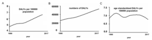
Figure 2. Changes in global burden of Age-related Macular Degeneration (AMD) in terms of disability-adjusted life years (DALY) numbers (A), DALY rates (B), and age-standardized DALY rates (C) between 1990 and 2017. Lines represent 95% uncertainty intervals [2]
Pharmacodynamic Evaluation Model for AMD Drugs
The in vivo pharmacology team at WuXi Biology uses sodium iodate (NaIO3)-induced retinal injury in mouse and monkey models to simulate dry AMD. NaIO3, an oxidizing agent, specifically targets the retinal pigment epithelium, mimicking key clinical aspects of dry AMD during model progression. This model facilitates the assessment of drug efficacy for dry AMD across various targets. Representative results are presented below:

Figure 3. Dose-dependent and time-dependent loss of photoreceptors induced by NaIO3 in C57BL/6J mice. After injecting multiple doses of NaIO3 (20, 35, and 50 mg/kg), retinal structure imaging was performed using Spectral Domain Optical Coherence Tomography (SD-OCT) at 1, 3and 21 days (Top). Retinal photoreceptor layer thickness (Bottom) was measured from the SD-OCT images. The results indicate a dose-dependent and time-dependent photoreceptor loss compared to the control group, with similar damage observed at 35 mg/kg and 50 mg/kg doses.
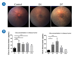
Figure 4. (A) Fundus deposition was observed in mice treated with 35 mg/kg NaIO3. From left to right: Fundus photographs of the control group and mice at 1 and 7 days post-NaIO3 treatment. Significant deposition (high reflectivity) was evident in the fundus. (B) Time-dependent changes in fundus complement levels after 35 mg/kg NaIO3 treatment. Left: Expression levels of complement 3a (C3a) over time. Right: Expression levels of complement 5a (C5a) over time. During the early stages post-NaIO3 injection, both C3a and C5a levels were significantly elevated, gradually returning to normal levels after day 7. *p<0.05, **p<0.01, ***p<0.001.

Figure 5. Impact of 35 mg/kg NaIO3 on photoreceptor response to light. Full-field flash electroretinogram (ffERG) responses under different light intensities (0.01 cd/m2/s and 3 cd/m2/s) were recorded at 1, 7, and 14 days post- NaIO3 injection (Left). Compared to the control group, NaIO3-treated mice showed a significant reduction in b-wave amplitude under both light conditions (top right for 0.01 cd/m2/s and bottom right for 3 cd/m2/s), indicating notable photoreceptor degeneration.

Figure 6. Gradual loss of photoreceptor cells in NaIO3-treated mice over time. Retinal sections stained with hematoxylin and eosin (HE) (Top) were used to quantify the number of cell nuclei (Bottom) from day 1 to day 28 post- NaIO3 treatment. A significant reduction in photoreceptor cells was observed from day 1 onwards.
Ophthalmic pharmacodynamic platform of the In Vivo Pharmacology Unit at WuXi Biology
The Ophthalmic Pharmacodynamic Platform at WuXi Biology is dedicated to the research and therapeutic development of eye diseases. Backed by a professional ophthalmic service team and systematic project management processes, the platform is equipped with comprehensive ocular testing devices for both small and large animals, and offers an extensive pharmacodynamics evaluation system, providing both morphological and functional assessments.
The team has validated over 30 ocular disease models, covering a wide range of eye diseases. They provide systematic ophthalmic efficacy evaluations, non-GLP safety assessments, and pharmacokinetic (PK) analyses. Additionally, the team has developed unique drug efficacy testing methodologies, offering end-to-end drug efficacy assessments from early screening to IND submission, thereby enabling the development of first-in-class drugs.
References
- Burton, M. J., Ramke, J., Marques, A. P., Bourne, R. R., Congdon, N., Jones, I., Ah Tong, B. A., Arunga, S., Bachani, D., Bascaran, C., Bastawrous, A., Blanchet, K., Braithwaite, T., Buchan, J. C., Cairns, J., Cama, A., Chagunda, M., Chuluunkhuu, C., Cooper, A., … Faal, H. B. (2021). The Lancet Global Health Commission on Global Eye Health: Vision Beyond 2020. The Lancet Global Health, 9(4).
- Xu, X., Wu, J., Yu, X., Tang, Y., Tang, X., & Shentu, X. (2020a). Regional differences in the global burden of age-related macular degeneration. BMC Public Health, 20(1).
WuXi AppTec | Disease Models:
- Read more about our in vivo pharmacology platform by clicking HERE
Related Content
Metabolic dysfunction-associated steatohepatitis (MASH) remains a significant global health challenge, driven by a complex interplay of lipid dysregulation, inflammation, and...
VIEW RESOURCESmall cell lung cancer (SCLC) remains one of the most aggressive and difficult-to-treat cancers, highlighting the ongoing need for new...
VIEW RESOURCE