
Endometriosis: Animal Models as Tools to Accelerate New Therapeutics
Introduction
Endometriosis is a common gynecological condition that significantly impacts the health of women of reproductive age worldwide. Approximately 10% of reproductive-aged women and girls are affected by this condition [1]. As a complex estrogen-dependent disease, endometriosis can onset from menarche and persist until menopause. There is currently no cure; treatments primarily aim to alleviate symptoms. Non-steroidal anti-inflammatory drugs (NSAIDs), progestins, and gonadotropin-releasing hormone (GnRH) analogs are commonly used to reduce pain and inflammation in patients. In the realm of new drug development, various novel analgesic and anti-inflammatory medications have shown potential therapeutic value. Animal models of endometriosis have become crucial tools in advancing the development of these new treatments.
Clinical Features and Pathogenesis of Endometriosis
Endometriosis is a chronic condition where tissue similar to the lining inside the uterus starts growing outside of it. This can lead to a variety of symptoms that significantly impact daily life, including intense menstrual cramps, pain during intercourse, pain during bowel movements and urination, chronic pelvic pain, bloating, nausea, fatigue, and even depression, anxiety, and infertility [2]. Moreover, endometriosis can cause broader health issues, such as affecting liver and fat metabolism, triggering systemic inflammation, and changing the expression of genes related to pain and mood in the brain.
The growth of this tissue varies greatly and can look different depending on where it is located. It can be classified into three main types: superficial peritoneal lesions, ovarian cysts known as endometriomas, and deeply infiltrating endometriosis (DIE). DIE refers to tissue that grows more than 5 mm deep into the peritoneum or into the walls of organs like the rectum and bladder. Interestingly, endometriosis is not limited to the pelvic area; it has been found in places like the diaphragm and lungs as well. This misplaced tissue responds to hormonal changes during the menstrual cycle, thickening, shedding, and bleeding just like the uterus lining, which can lead to inflammation and scarring in nearby areas. Estrogen is a key player in promoting the growth of this tissue. While many women see improvement in their symptoms after menopause when estrogen levels drop, this is not the case for everyone.

Figure 1. Common Symptoms of Endometriosis [3]
Endometriosis is a complicated disease whose exact cause is still not fully understood. Multiple factors could contribute to its onset and progression. The most widely accepted explanation is Sampson’s theory of retrograde menstruation [4]. This theory suggests that during menstruation, some menstrual blood containing endometrial-like cells flows backward through the fallopian tubes into the pelvic cavity, where these cells implant and grow outside the uterus. While substantial evidence supports this theory, retrograde menstruation is not the only pathway for developing endometriosis. Recent research has discovered that stem cells from bone marrow or other sources can travel through blood and lymphatic vessels, differentiating into endometrial-like cells [5]. The cellular metaplasia hypothesis proposes that non-endometrial cells from locations like the peritoneum, pleura, and ovaries can transform into endometrial-like cells under certain conditions [6]. Besides these factors, abnormal immune function and genetic predisposition are also believed to play significant roles in the development and progression of endometriosis.
Diagnosis and Treatment of Endometriosis
Endometriosis shares symptoms with several other gynecological conditions, such as primary dysmenorrhea, ovarian cysts, pelvic inflammatory disease, adenomyosis, and uterine fibroids. Endometriosis also has overlapping symptoms with some bladder and musculoskeletal disorders. This symptom similarity can lead to delayed diagnosis and misdiagnosis of endometriosis. While histological examination is crucial for a definitive diagnosis, in practice, imaging tests are often more commonly used because the lesions can be very small or deeply embedded in tissues. Transvaginal ultrasound can help identify ovarian cysts and other pelvic abnormalities. Pelvic MRI provides detailed information about the tissue structures, including the exact location, size, and relationship of the lesions to surrounding tissues, which supports both diagnosis and treatment planning.
Currently, there is no cure for endometriosis. The main goals of treatment are to relieve pain, control the growth of the lesions, improve quality of life, and enhance fertility. Common treatment methods include medication and surgery:
Medications:
- Non-Steroidal Anti-Inflammatory Drugs (NSAIDs): These medications are primarily used to relieve pain and inflammation associated with endometriosis. NSAIDs can significantly improve pain symptoms for many patients, but they typically do not have a direct effect on the growth of endometrial lesions.
- Hormonal Therapies: This category includes oral contraceptives, progestins, and GnRH analogs. These medications work by regulating hormone levels to inhibit the growth of endometrial-like tissue, thereby alleviating pain symptoms.
Surgery:
For patients who do not respond well to medication, surgery is another treatment option. The main types of surgery include laparoscopy and laparotomy, with the choice depending on the extent and severity of the lesions. Surgical removal of ectopic endometrial tissue can help restore the normal structure of the pelvic cavity. For those experiencing infertility due to endometriosis, assisted reproductive technology, such as in vitro fertilization, can be utilized to achieve pregnancy. These advanced techniques offer hope for many women wishing to conceive despite the challenges posed by endometriosis.
Preclinical Animal Models of Endometriosis
Animal models are crucial tools for studying the pathological mechanisms of endometriosis and testing new drugs. Commonly used models include mice, rats, and non-human primates (NHP) [7]. Although the reproductive systems of rodents differ from humans, their physiological and anatomical similarities are sufficient to replicate many pathological features of endometriosis. Rodent models of endometriosis are well-established, with standardized techniques that ensure high reproducibility and comparability of experimental results. Unlike humans, rodents do not experience menstrual bleeding, so endometriosis cannot be induced through retrograde menstruation. Instead, these animals have short estrous cycles, typically lasting 4 to 5 days. To mimic human endometriosis, autologous or heterologous endometrial tissue is transplanted onto the abdominal wall or injected into the peritoneal cavity of rodents. This method effectively simulates the formation of endometriotic lesions, providing valuable insights into the disease and potential treatments.
Case Study:
The Urogenital and Endocrine Diseases team of WuXi Biology has observed significant findings in a mouse model of endometriosis. By transplanting allogeneic mouse endometrial tissue onto the abdominal wall, we observed that the transplanted tissue gradually grew (Figures 2D, E) and induced noticeable pain hypersensitivity in the animals (Figures 2B, C). When endometrial fragments were injected into the mouse peritoneal cavity, these fragments adhered to locations such as the abdominal wall, mesentery, and fat, developing into lesion tissue and causing pain (Figure 3). Both modeling methods demonstrated similar phenotypes to clinical endometriosis, including ectopic tissue growth, pain, and inflammation. Moreover, treatment with the GnRH analog, cetrorelix acetate, significantly improved both lesion growth and pain phenotypes (Figures 2, 3).
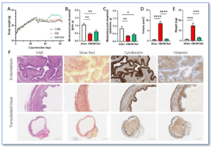
Figure 2. Mouse Endometriosis Transplantation Model (Source: Internal data from WuXi Biology) A. Body weight changes; B. Hind paw mechanical pain threshold; C. Abdominal mechanical pain threshold; D. Transplanted tissue volume; E. Transplanted tissue weight; F. Histopathology of transplanted tissue
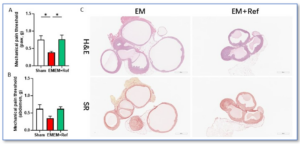
Figure 3. Mouse Endometriosis Infusion Model (Source: Internal Data from WuXi Biology) A. Hind paw mechanical pain threshold; B. Abdominal mechanical pain threshold; C. Histopathology of ectopic tissue
Conclusion
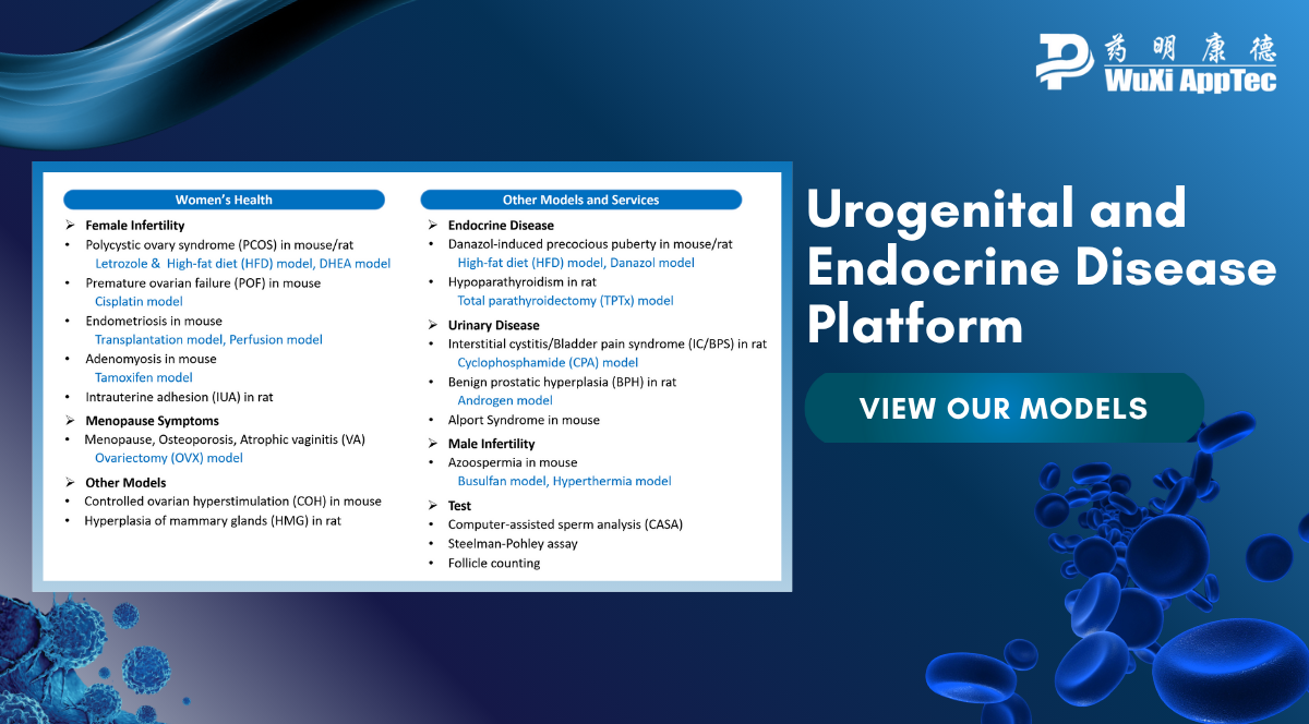
Endometriosis is a prevalent and complex chronic gynecological disease that significantly impacts women’s health. Although its exact cause remains unclear, extensive research has been conducted on its symptoms, diagnosis, and treatment options. The Urogenital and Endocrine Diseases team of WuXi Biology is dedicated to addressing various women’s health issues, including endometriosis, and has successfully developed multiple disease models (Figure 4). Reliable preclinical evaluation models and strategies are essential for advancing drug development for endometriosis. These models help drive the progress of new therapies in the field of women’s health, contributing to the improvement of overall female well-being.
Figure 4. Urogenital and Endocrine Disease Model Platform of WuXi Biology: Models and Services
References:
- World Health Organization (WHO): Endometriosis. 24 March 2023. https://www.who.int/news-room/fact-sheets/detail/endometriosis
- Taylor HS, Kotlyar AM, Flores VA. Endometriosis is a chronic systemic disease: clinical challenges and novel innovations. Lancet. 2021. 397(10276):839-852. doi: 10.1016/S0140-6736(21)00389-5
- Saunders PTK, Horne AW. Endometriosis: Etiology, pathobiology, and therapeutic prospects. Cell. 2021. 184(11):2807-2824. doi: 10.1016/j.cell.2021.04.041.
- Sampson, J.A. (1927) Peritoneal Endometriosis Due to the Menstrual Dissemination of Endometrial Tissue into the Peritoneal Cavity. American Journal of Obstetrics & Gynecology, 14, 442-469.
- Du, H., & Taylor, H. S. (2007). Contribution of bone marrow-derived stem cells to endometrium and endometriosis. Stem cells (Dayton, Ohio), 25(8), 2082–2086.
- Clement P. B. (2007). The pathology of endometriosis: a survey of the many faces of a common disease emphasizing diagnostic pitfalls and unusual and newly appreciated aspects. Advances in anatomic pathology, 14(4), 241–260.
- Endometriosis in the Mouse: Challenges and Progress Toward a ‘Best Fit’ Murine Model. Frontiers in Physiology. 2022. 12:806574. doi: 10.3389/fphys.2021.806574. eCollection 2021.
Related Content
Strategies for Novel Autoimmune Drug Development: Preclinical Efficacy and Case Studies of JAK-STAT Targeted Therapies JAK Family Molecules: Central Hubs...
VIEW RESOURCEAccelerating SLE Drug Discovery: A Short-Term BM12-Induced Mouse Model for Efficient Drug Screening Systemic lupus erythematosus (SLE) is a chronic...
VIEW RESOURCE