
Brain metastasis mouse models for the evaluation of multikinase inhibitors on ROS1-fusion-positive lung cancer
Lung cancer is the leading cause of cancer-related death worldwide. Nearly 80% of lung cancers are non-small cell lung cancer (NSCLC) and 60% of them are diagnosed at the metastatic stage. Brain metastases affect more than 20% of NSCLC patients with poor prognosis and disabling symptoms. However, few therapies have been approved for the treatment of lung cancer brain metastases. A panel of rapid, predictive and clinically-relevant animal models are urgently needed to study the biology of brain metastases and to identify effective therapeutic approaches.
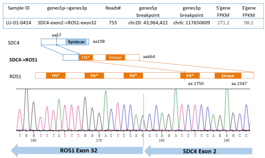
Here we describe methods for efficient establishment of brain metastases mouse models via different injection route (intracranial or intracarotid). We established a novel ROS1 positive patient-derived xenograft (PDX) model together with its PDC sub-line, in which exon 2 of SDC4 was fused to exon 32 of ROS1 (SDC4ex2-ROS1ex32). Cells have been transduced with luciferase expression vector for in vivo imaging detection. Three ROS1 inhibitors were tested on these brain metastasis models to compare their efficacies.
In summary, a ROS1 positive patient-derived xenograft model together with its PDC-luc subline for in-vivo brain metastasis model was established. In-vivo brain metastases models were established via intracranial and intracarotid injection to provides a reliable platform for identifying the mechanisms of metastatic development as well as a clinically relevant therapeutic screening.
2 Brain metastasis ROS1 model_5989_Hui
Related Content
KRAS G12D inhibitors continue to advance, but treatment resistance remains an important challenge. Understanding how that resistance develops is critical...
VIEW RESOURCEMetabolic dysfunction-associated steatohepatitis (MASH) remains a significant global health challenge, driven by a complex interplay of lipid dysregulation, inflammation, and...
VIEW RESOURCE